10. Vacuole 11. Hyaloplasm 12. Lysosome 13. Centrosome (Centriole)
eukaryotes, or Nuclear(lat. Eucaryota from the Greek εύ- - good and κάρυον - nucleus) - the kingdom of living organisms, whose cells contain nuclei. All organisms except bacteria and archaea are nuclear.
The structure of a eukaryotic cellEukaryotic cells are, on average, much larger than prokaryotic cells, the difference in volume reaches thousands of times. Eukaryotic cells include about a dozen different types of structures known as organelles (or organelles, which, however, somewhat distorts the original meaning of the term), of which many are separated from the cytoplasm by one or more membranes. In prokaryotic cells, there is always a cell membrane, ribosomes (significantly different from eukaryotic ribosomes) and genetic material - a bacterial chromosome, or genophore, but membrane-enclosed internal organelles are rare. The nucleus is the part of the cell surrounded by a double membrane (two elementary membranes) in eukaryotes and containing genetic material: DNA molecules "packed" into chromosomes. The nucleus is usually one, but there are also multinucleated cells. Division into kingdomsThere are several options for dividing the superkingdom of eukaryotes into kingdoms. The kingdoms of plants and animals were the first to be distinguished. Then the kingdom of fungi was singled out, which, due to biochemical characteristics, according to most biologists, cannot be assigned to any of these kingdoms. Also, some authors distinguish the kingdoms of protozoa, mixomycetes, chromists. Some systems have up to 20 kingdoms. Differences between eukaryotes and prokaryotesThe most important, fundamental feature of eukaryotic cells is associated with the location of the genetic apparatus in the cell. The genetic apparatus of all eukaryotes is located in the nucleus and is protected by a nuclear membrane (in Greek, "eukaryote" means having a nucleus). Eukaryotic DNA is linear (in prokaryotes, DNA is circular and floats freely in the cytoplasm). It is associated with histone proteins and other chromosomal proteins that bacteria do not have. In the life cycle of eukaryotes, there are usually two nuclear phases (haplophase and diplophase). The first phase is characterized by a haploid (single) set of chromosomes, then, merging, two haploid cells (or two nuclei) form a diploid cell (nucleus) containing a double (diploid) set of chromosomes. After a few divisions, the cell becomes haploid again. Such a life cycle and, in general, diploidy are not characteristic of prokaryotes. The third, perhaps the most interesting, difference is the presence of special organelles in eukaryotic cells that have their own genetic apparatus, multiply by division and are surrounded by a membrane. These organelles are mitochondria and plastids. In their structure and activity, they are strikingly similar to bacteria. This circumstance prompted modern scientists to the idea that such organisms are descendants of bacteria that have entered into a symbiotic relationship with eukaryotes. Prokaryotes are characterized by a small number of organelles, and none of them is surrounded by a double membrane. In prokaryotic cells, there is no endoplasmic reticulum, Golgi apparatus, or lysosomes. It is equally important, describing the differences between prokaryotes and eukaryotes, to say about such a phenomenon in eukaryotic cells as phagocytosis. Phagocytosis (literally "eating") is the ability of eukaryotic cells to capture and digest a variety of solid particles. This process provides an important protective function in the body. It was first discovered by I.I. Mechnikov near starfish. The appearance of phagocytosis in eukaryotes is most likely associated with average sizes (more on size differences below). The size of prokaryotic cells is incommensurably smaller, and therefore, in the process of evolutionary development, eukaryotes faced the problem of supplying the body with a large amount of food, as a result, the first predators appear in the eukaryotic group. Most bacteria have a cell wall that is different from the eukaryotic one (not all eukaryotes have it). In prokaryotes, it is a strong structure composed mainly of murein. The structure of murein is such that each cell is surrounded by a special mesh bag, which is one huge molecule. Among eukaryotes, fungi and plants have cell walls. In fungi, it consists of chitin and glucans, in lower plants from cellulose and glycoproteins, diatoms synthesize a cell wall from silicic acids, in higher plants from cellulose, hemicellulose and pectin. Apparently, for larger eukaryotic cells, it has become impossible to create a cell wall of high strength from a single molecule. This circumstance could force eukaryotes to use a different material for the cell wall. The metabolism of bacteria is also varied. In general, there are four types of nutrition, and all of them are found among bacteria. These are photoautotrophic, photoheterotrophic, chemoautotrophic, chemoheterotrophic (phototrophic use the energy of sunlight, chemotrophic use chemical energy). Eukaryotes, on the other hand, either synthesize energy from sunlight themselves, or use ready-made energy of this origin. This may be due to the appearance of predators among eukaryotes, the need to synthesize energy for which has disappeared. Another difference is the structure of the flagella. In bacteria, they are thin - only 15-20 nm in diameter. These are hollow filaments made from flagellin protein. The structure of eukaryotic flagella is much more complicated. They are a cell outgrowth surrounded by a membrane and contain a cytoskeleton (axoneme) of nine pairs of peripheral microtubules and two microtubules in the center. In contrast to rotating prokaryotic flagella, eukaryotic flagella bend or wriggle. The two groups of organisms we are considering, as already mentioned, differ greatly in their average size. The diameter of a prokaryotic cell is usually 0.5-10 microns, while the same indicator in eukaryotes is 10-100 microns. The volume of such a cell is 1000-10000 times greater than that of a prokaryotic cell. In prokaryotes, ribosomes are small (70S-type). Eukaryotes have larger ribosomes (80S-type). Apparently, the time of occurrence of these groups also differs. The first prokaryotes arose in the process of evolution about 3.5 billion years ago, and eukaryotic organisms originated from them about 1.2 billion years ago. |
1. The diversity of organisms on Earth, the similarity of their structure and life:
cellular structure, similar structure of cells, similarity of chemical composition,
metabolism, reproduction.
2. Differences in cell structure are the basis for dividing all organisms into two large groups: pre-nuclear (prokaryotes) and nuclear (eukaryotes). Examples of pre-nuclear organisms: bacteria and blue-green algae.
Examples of nuclear organisms: humans, animals, plants, fungi.
3. Features of the structure of pre-nuclear organisms: 1)
absence of a formed nucleus, nuclear membrane, nuclear substance is located
in the cytoplasm; 2) DNA is concentrated in one chromosome, which has the shape of a ring and
located in the cytoplasm; 3) the absence of a number of organelles: mitochondria,
endoplasmic reticulum, Golgi apparatus; 4) all organisms of this group
unicellular.
4. Cell of non-nuclear organisms, such as bacteria,
has a dense shell of carbohydrates, plasma membrane, nuclear substance
(chromosome), cytoplasm, very small ribosomes.
5. Features of the structure of nuclear organisms: 1) the presence
in a cell of a formed nucleus, delimited from the cytoplasm by a membrane with pores; 2)
the presence of the entire complex of cytoplasmic organelles: mitochondria, the Golgi apparatus,
lysosomes, ribosomes, endoplasmic reticulum, cell center, as well as
plasma membrane and outer shell in plant cells, fungi; 3)
the presence of several chromosomes located in the nucleus.
6. Diversity of nuclear organisms by structure
(unicellular and multicellular), according to the method of nutrition (autotrophs, heterotrophs,
vegetative).
2. Biological diversity, its role in maintaining the sustainability of the biosphere.
I. Biodiversity - the variety of species that inhabit the Earth, the diversity
natural ecosystems on the globe.
2. The diversity of species in nature is the reason for the various food, territorial relationships between them, the most complete use of natural resources, and the closed circulation of substances in the natural ecosystem. The rainforest is a stable ecosystem due to the wide variety of species in it, the adaptability of organisms to cohabitation, and the optimal use of natural resources. An ecosystem consisting of a small number of species, such as a small reservoir, a meadow, is an example of unstable natural communities.
3. Reduction of species diversity as a result of activities
human: the construction of cities, railways and highways, cutting down large
forests, construction of industrial enterprises, plowing of land for
farmland. Extinction is currently about 10% of the species
higher plants on earth. Deforestation of tropical forests, in which
a significant part of plant and animal species is a problem that requires the use
special forest protection measures. Extinction over the past 400 years of more than 60 species
mammals and over 100 bird species.
4. The impact of environmental pollution on species diversity, the reasons for its reduction. Thus, water pollution in rivers by industrial waste is the reason for the reduction in the number of crayfish, freshwater pearl mussel (mollusk), and some species of fish. The treatment of fields and gardens with pesticides is the cause of the death of birds that feed on insects infected with poisons. Ecosystem-dark nature of the decline in species diversity: each extinct plant species takes with it five species of invertebrates
animals whose existence is inextricably linked with this plant.
5. The role of biodiversity in maintaining the stability of the biosphere. The dependence of human existence on the state of the biosphere, on its biological diversity. Conservation of species diversity, habitats of plants and animals. Protected areas: nature reserves, biosphere reserves, national parks, natural monuments, their role in conservation
diversity of life on earth.
TICKET#13
Biospheric prerequisites for the emergence of eukaryotic organisms
Vladimir Malakhov
One hundred years ago, the Russian biologist K.S. Merezhkovsky suggested that the eukaryotic cell arose as a result of the symbiosis of several independent organisms. This idea has become one of the main paradigms of modern biology.
All living organisms that inhabit our planet are divided into two large groups: prokaryotes (non-nuclear) and eukaryotes (nuclear). Prokaryotes are bacteria whose hereditary material is represented by a simple circular DNA molecule. Nuclear are various unicellular and multicellular organisms (protozoa, plants, animals and fungi), in the cells of which there is a formed nucleus with chromosomes in which linear DNA molecules are associated with special nuclear proteins - histones. In addition to the nucleus in the cells of eukaryotic organisms, there are other organelles: mitochondria, flagella, chloroplasts. When and how did the eukaryotic organisms that dominate the modern biosphere originate?
According to modern concepts, our planet was formed about 4.5 billion years ago. Initially, the Earth was dry, water appeared as a result of degassing of the bowels - the release of water vapor and gases into the atmosphere, which made up the ancient atmosphere. As the water vapor condensed, small puddles first appeared, which gradually became larger and larger. However, it took 500-700 million years for more or less large water bodies to appear on Earth, which gradually formed the hydrosphere - the liquid shell of our planet, which currently occupies about 70% of its surface. Then, as a result of sedimentation of various particles to the bottom of reservoirs, sedimentary rocks were formed.
The oldest sedimentary rocks are graphitized shales from the Isua Formation in Greenland - their age is about 3.8 billion years. It is surprising that in these rocks undoubted signs of life that once existed were found - traces of the activity of organisms that carried out the process of photosynthesis. The fact is that in organic matter created in the process of photosynthesis, the ratio of carbon isotopes 12C and 13C changes in favor of the lighter 12C isotope. And no matter what happens to this substance in the future, this ratio will be preserved in it. The carbon in the Isua Shale is clearly of organic origin. This means that already 3.8 billion years ago in the primary water bodies of the planet (most likely the World Ocean did not yet exist at that time) organisms capable of photosynthesis lived. Fossilized cells similar to modern cyanobacteria have been found in rocks 3.5 billion years old (Warravuna Formation in Australia). Slightly younger deposits (more than 3.1 billion years) contain the remains of chlorophyll - phytane and pristane, as well as specific pigments of cyanobacteria - phycobilins.
Of course, among the organisms of that time were not only photosynthetics, using the energy of sunlight, but also chemosynthetics, receiving energy through various chemical reactions. In the first billion years of the existence of the biosphere, due to the activity of chemosynthetic bacteria, many (if not most) of the ore deposits that mankind still uses were formed, therefore fossilized remains of bacteria are often found in ore bodies. For example, such a large deposit of iron ore as the Kursk magnetic anomaly, according to modern data, was formed as a result of the activity of bacteria.
There is no doubt that for a significant part of its history (at least 2 billion years), the biosphere was prokaryotic, that is, it included only organisms similar to modern bacteria. Eukaryotic organisms - a variety of unicellular protozoa, and later (600-800 million years ago) and multicellular organisms - took their place in the biosphere only about 1 billion years ago.
Prokaryotes and eukaryotes are the two main species of living beings on our planet. Biologists and physicians, however, are actively studying another group of biological objects - viruses, but they exhibit the properties of a living organism only inside the cells of their "masters". The sizes of prokaryotic cells in most cases range from 0.5 to 3 microns, and the smallest (mycoplasmas) do not exceed 0.10-0.15 microns. Giant cells of some sulfur bacteria reach 100 microns in length, and spirochete cells sometimes grow up to 250 microns. The main feature of prokaryotes is the absence of a nucleus. Their genetic material (genophore) is represented by a single circular double-stranded DNA molecule attached to the cytoplasmic membrane that dresses the cell. Prokaryotes do not have a nuclear membrane, an endoplasmic reticulum (sometimes there are protrusions of the surface membrane - the so-called mesosomes), mitochondria, plastids and other cytoplasmic organelles characteristic of eukaryotes. They also lack microtubules, so they have neither centrioles nor spindles. Ribosomes of prokaryotes lack one of the types of ribosomal RNA (the so-called 5.8S RNA) and have a smaller mass than that of eukaryotes. Usually, the mass of ribosomes is estimated by the so-called sedimentation constant (an indicator of the sedimentation rate during centrifugation). For prokaryotic ribosomes, it is 70S, and for eukaryotes, it is 80S.
Prokaryotes have a huge (compared to eukaryotes) variety of metabolic processes. They are capable of fixing carbon dioxide, nitrogen, various types of fermentation, oxidation of various inorganic substrates (compounds of sulfur, iron, manganese, nitrites, ammonia, hydrogen, etc.). There are many photosynthetic forms among prokaryotes, first of all, cyanobacteria that are often found in the modern biosphere, which are also called blue-green algae. They (or related organisms) were widespread in the distant past. Geological structures created by ancient cyanobacteria (probably together with other photosynthetic prokaryotes) - stromatolites - are often found in the oldest layers of the earth's crust, corresponding to the Archean and early Proterozoic. The activity of photosynthetic and other autotrophic prokaryotes, which began about 4 billion years ago, had several important consequences.
The first has to do with changes in the Earth's atmosphere. The fact is that in ancient times it was practically anoxic. As a result of photosynthesis, molecular oxygen began to be released into the atmosphere, but quickly associated with non-oxidized components of the lithosphere - iron and other metals. Therefore, despite the presence of a constant source of free oxygen, the biosphere remained predominantly anaerobic. Living organisms during this period were also represented mainly by anaerobes. In the meantime, banded iron ores (the so-called jaspilites) were deposited in the lithosphere, in which oxidized iron alternated with underoxidized. Under anoxic conditions, pyrites (ores of the FeS2 type) were deposited, which could not be formed in the presence of free oxygen. Findings of such fossils make it possible to establish that, despite the abundance of photosynthetics, the anaerobic period in the development of the biosphere lasted almost 2 billion years.
However, about 2 billion years ago, the oxygen content in the atmosphere reached 1% and continued to rise, since by that time most of the iron and other metals on the surface had been oxidized. At the same time, the amount of iron and other metals rising from the depths of the Earth gradually decreased. During the formation of the planet, heavy and light components were randomly mixed. Later, in the process of gravitational differentiation, the metals gradually sank to the center of the planet, forming its iron core, and the light components - silicates - rose up, forming the mantle.
For anaerobic organisms, the increase in oxygen concentration was a disaster, since oxygen is a very aggressive element, it quickly oxidizes and destroys organic compounds. If in the anaerobic biosphere, in the thickness of the stromatolites, aerobic pockets remained, from where the oxygen accumulated as a result of photosynthesis diffused into the atmosphere, now the biosphere, according to the apt expression of Academician G.A. Zavarzin, "turned inside out" - it turned into an oxygen one with a few oxygen-free pockets, where anaerobic microorganisms found shelter. In the new aerobic atmosphere, only those few prokaryotes (oxybacteria) could survive, which even earlier in oxygen pockets in the thickness of stromatolites adapted to high oxygen concentrations.
The second important consequence of the activity of autotrophic prokaryotes is the accumulation of deposits of organic matter. The biotic cycle of substances in the biosphere, consisting exclusively of prokaryotes, was very imperfect. The biomass created by autotrophic bacteria was decomposed mainly under the influence of abiotic physical and chemical processes in the environment. Undoubtedly, heterotrophic bacteria also played a significant role in the decomposition of the biomass created by prokaryotic autotrophs, but their capabilities were limited due to the peculiarities of the organization of prokaryotic cells. As you know, prokaryotes are fundamentally incapable of swallowing their victims. Predation in bacteria is very rare and looks very unusual. The predatory bacterium Bdellovibrio is much smaller than its victims, it penetrates the cell wall of the bacterium and multiplies inside the body of the unfortunate. 
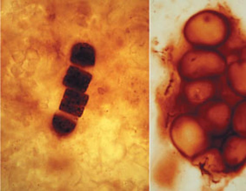
Cells of fossil prokaryotic organisms close to cyanobacteria in thin sections of Archean sedimentary rocks (left and center). On the right is a photograph of fossil stromatolites formed by ancient photosynthetic bacteria.
Why are prokaryotes incapable of swallowing food? The fact is that they lack actin and myosin - proteins that ensure the mobility of the cytoplasm in eukaryotes. Thanks to them, during the capture of food particles (phagocytosis) and the formation of digestive vacuoles, pseudopodia are formed (temporary cytoplasmic outgrowths that serve to move and capture food). Prokaryotes cannot do this. Heterotrophic bacteria secrete enzymes into the external environment, a kind of "external digestion" (exofermentation) occurs, and low molecular weight products are absorbed through the cytoplasmic membrane. All this led to a low rate of biomass decomposition created by autotrophic prokaryotes. Therefore, in the early stages of the evolution of the biosphere, huge masses of organic carbon were removed from the biological cycle, preserved in sediment, subjected to chemical transformation, turning into oil shale, oil and gas, which mankind is still actively using.
Only the appearance of microscopic aerobic predators that would swallow bacteria, digest them and return carbon (preferably in the form of CO2), nitrogen (in the form of ammonium compounds), phosphorus and others to the biosphere could improve the biological cycle, accelerate the return of carbon and other nutrients into it. biogenic elements. The first eukaryotic organisms became such predators.
Predators
Eukaryotes have two universal proteins - actin and myosin, which provide various types of cellular mobility: amoeboid activity, movement of organelles inside the cell, and in higher organisms - muscle contractions. The actin-myosin system allows the formation of pseudopodia, capturing the victim with them and forming digestive vacuoles (even viruses enter the eukaryotic cell by provoking the so-called "endocytosis" - the cell takes them for something useful, "swallows", and the virus, once in the cytoplasm, begins its destructive work). The acquisition of the actin-myosin system allowed eukaryotes to feed by phagocytosis, actively capturing large food particles.
The emergence of such organisms has accelerated the biotic cycle to an extraordinary degree, as they have become consumers of bacterial biomass. Digesting bacterial cells, phagotrophic eukaryotes quickly returned elements to the cycle of substances, which before that could again enter it only through slow decomposition. It can be assumed that the appearance of eukaryotes led to a sharp decrease in "bacterial fossils", that is, deposits of organic and inorganic substances resulting from the activity of bacteria.
The ability of eukaryotes to capture food particles meant that the predator must be larger than the prey. Indeed, the linear dimensions of small soil amoebae or flagellates are approximately 10 times the size of the bacteria they feed on. Thus, the volume of the cytoplasm of eukaryotes is about 1000 times larger than that of prokaryotes, which also requires a large number of gene copies to supply the cytoplasm with transcription products. One way to solve this problem is to increase the number of genophores, that is, circular DNA molecules. Large (so-called "polyploid") bacteria and eukaryotic ancestors with a large volume of cytoplasm followed this path. Multiple genophores (originally identical) became the rudiments of chromosomes, in which differences gradually accumulated. 
Cyanobacteria also thrive in the modern biosphere (left photo). In some places they form structures resembling ancient stromatolites. The right photo shows modern stromatolites from Shark Bay in northern Australia. With amoeboid movement and nutrition by phagocytosis, the cytoplasm of the cell (especially the peripheral one) becomes very mobile. The genophores attached to the surface membrane of the cell found themselves in the zone of strong cytoplasmic currents; therefore, a membrane-protected area appeared in the central cytoplasm, where the genophores were stored. The process could occur in different ways, but one of the possible ways is deep invaginations of sections of the cytoplasmic membrane with genophores attached to them (after all, the nuclear envelope is part of the endoplasmic reticulum of a eukaryotic cell, which can be associated with the external environment).
Thus, primary eukaryotes had a nucleus limited by a double nuclear membrane - a derivative of the endoplasmic reticulum, but also had a ring structure of genophores and were deprived of specific nuclear proteins - histones. Surprisingly, a similar structure of the nucleus has been preserved in some modern eukaryotes, for example, in dinoflagellates. In these protozoa, the nucleus is surrounded by a double nuclear membrane, but the chromosomes contain circular DNA molecules devoid of histones. Apparently, the core of dino-flagellates is a relic structure that has retained the structure characteristic of primary eukaryotic organisms.
Symbiotic origin of mitochondria and flagella
The ability to phagotrophic nutrition predetermined the possibility of the appearance of intracellular symbionts in eukaryotes. Prokaryotes could not do this - deprived of the ability to swallow anyone, they did not acquire intracellular endosymbionts. For eukaryotes, on the contrary, the inclusion of various prokaryotic and eukaryotic organisms as intracellular symbionts is quite typical. The eukaryotic cell arose as a result of the symbiosis of the primary amoeboid organism with various prokaryotic and eukaryotic creatures. This provision formed the basis of the so-called concept of symbiogenesis, which has become one of the paradigms of modern biology.
 |
 |
| "Predation" in modern prokaryotes. Above, the "predatory" bacterium Bdellovibrio enters E. coli and multiplies inside it. Below, a bacteriophage virus injects its DNA into the bacterium, while its protein shell remains outside. Viral DNA provides the synthesis of new viral particles. | Through the actin-myosin system, eukaryotic organisms can form pseudopodia and phagocytize bacteria and other particles (top). The virus uses this property of eukaryotic organisms and provokes endocytosis - the absorption of a viral particle by the cell itself (below). |
According to current ideas, such important organelles of the eukaryotic cell as mitochondria have a symbiotic origin. They provide the synthesis of the main energy resource of any cell - ATP due to oxidative phosphorylation, which is possible only in the presence of oxygen. Only some protozoa that live in anaerobic conditions (for example, in the intestines of animals or in oxygen-deprived swamp waters) do not have mitochondria. Undoubtedly, the absence of mitochondria in them is a secondary sign associated with existence in anoxic conditions, this is confirmed by the fact that some mitochondrial genes were found in the genome of such protozoa.
As is known, mitochondria are surrounded by two membranes, the inner one (the one that forms mitochondrial cristae) belongs to the mitochondrion itself, and the outer one belongs to the vacuole containing the symbiont. Mitochondria have their own hereditary material, organized in the same way as in prokaryotes. This is a histone-free circular DNA molecule that carries information about proteins that are synthesized in the mitochondria itself on its own prokaryotic-type ribosomes with a sedimentation constant of 70S. True, in mitochondria, the circular DNA molecule is about a hundred times shorter than in bacteria that exist independently. The fact is that many mitochondrial proteins are encoded in the nuclear DNA of eukaryotic cells. Apparently, in the process of long-term joint evolution of the host cell and the symbiont, a significant part of the genes from the mitochondrial genome passed into the nucleus of the eukaryotic cell. The mitochondrial genome contains genes for only those proteins that cannot overcome the two-membrane barrier (for example, hydrophilic proteins). However, mitochondria are not born again in the cell - they divide in the same way as free-living bacteria. What kind of prokaryotes could be the ancestors of mitochondria? Among modern prokaryotes, purple alpha-proteobacteria are closest to them (this is evidenced, in particular, by new data of molecular phylogeny) - aerobic photosynthetic bacteria, the membrane of which forms deep invaginations similar to mitochondrial cristae. The progenitors of such bacteria probably lived in the oxygen pockets of the anaerobic biosphere. Having entered into symbiosis with ancient amoeboid eukaryotes, proteobacteria lost the ability to photosynthesize, since they began to receive all the necessary organic substances from the host - an ancient eukaryote, who received his benefit: he ceased to be afraid of high concentrations of oxygen, which was utilized by symbionts.Primary aerobic eukaryotes with symbionts initially also populated oxygen pockets, but when the oxygen concentration began to increase 3 billion years after the formation of the biosphere, eukaryotes were able to spread widely in the biosphere. In the layers of the earth's crust related to this period, the so-called acritarchs appear - large spherical cells with a diameter of 50-60 microns. They could not belong to prokaryotes whose spherical cells do not exceed a few microns in diameter (filamentous forms can be much longer). In the layers, whose age is about 1.7 billion years, sterols, substances synthesized in the nucleus of eukaryotic organisms, were found. Thus, in the period from 1 to 2 billion years ago, the adaptive evolution of eukaryotes began.
The flagella are not separated from the cytoplasm by membranes, there are no obstacles for the transfer of proteins from the cytoplasm to the flagellum, so most of the flagellum proteins are encoded in the cell nucleus. At the same time, inside the basal body of the flagellum there is a small circular DNA molecule that contains several genes that control the formation of the basal body. The fact is that centrioles (basal bodies) do not appear in the cell from scratch. Before dividing, two centrioles diverge and a new one is formed next to each of them. Thus, for the synthesis of the next organoid, a "seed" in the form of the old one is needed.
It is assumed that the ancestors of the flagellum were bacteria resembling modern spirochetes, motile bacteria whose narrow, spirally twisted cells move rapidly, as if screwing into space. True, they themselves could not be the ancestors of flagella: they do not have microtubules, and the fine structure is completely different. But this does not mean at all that in the distant past there were no other spirochete-like organisms, which became the ancestor of the eukaryotic flagellum. Apparently, its progenitors were first exosymbionts, that is, attached to the cytoplasmic membrane of a primitive eukaryote from the outside. The symbiont used the metabolites secreted by the host for its nutrition, and in return, due to its locomotor activity, contributed to its rapid (compared to the formation of pseudopodia) movement. It is this interaction that has formed between spirochetes and some large protozoa. Symbiotic spirochetes sit on the surface of the flagellate Myxotricha paradoxa (which also has ordinary flagella), their movements are coordinated, like in real cilia, and locomotor activity provides a smooth and gradual movement of the flagellate, while its own flagella allow it to make only fast movements forward in a spiral. It is curious that for greater convenience of attachment of spirochetes, the host cell kindly forms special compacted "stands", from which bundles of fibrils extend into the host cytoplasm, resembling the roots of real flagella and cilia. This example shows that symbiosis between motile bacteria and eukaryotes can occur repeatedly.
Origin of eukaryotic plants
Primary eukaryotes were single-celled animals. They fed by capturing and digesting other microscopic organisms. One of the main directions of their evolution was the acquisition of photosynthetic symbionts, which turned into organelles that provided the synthesis of organic substances from carbon dioxide and water due to the energy of sunlight. This path led to the emergence of various groups of eukaryotic plants, that is, autotrophic photosynthetic organisms. They are not related to each other and arose as a result of the symbiosis of predatory protists (protozoa or their colonies) with various photosynthetic organisms.
In several cases, cyanobacteria became symbionts of predatory eukaryotes - blue-green algae, the most common (at least in the modern biosphere) and, perhaps, the oldest group of photosynthetic prokaryotes. Their undoubted descendants are the photosynthetic organelles (chloroplasts) of red algae. They are surrounded by only two membranes, have their own circular DNA and prokaryotic-type ribosomes, and contain chlorophyll "a" typical of cyanobacteria and specific pigments of cyanobacteria - phycobilins. Red algae are currently widespread in the seas of our planet. They are able to exist at depths of several hundred meters, but they also live in the tidal zone, and some species also live in fresh waters. Perhaps red algae are the oldest group of eukaryotic plants. This is evidenced by the complete absence of flagellated stages in their life cycle (even their spermatozoa are flagellaless), which suggests that the ancestors of these algae separated from the rest of the eukaryotes even before the acquisition of flagella.
However, red algae are not the only group using descendants of cyanobacteria as symbionts. In unicellular flagellates - glaucophytes (not at all related to red algae), photosynthetic organelles are called cyanella. They even retain the murein membrane characteristic of cyanobacteria (i.e., a mechanically strong element of the cell wall). Nevertheless, cyanella are true symbionts that cannot live separately from the host. Even their genome - circular DNA - is about 10 times shorter than that of free-living cyanobacteria. This means that in this case, too, a significant part of the cyanella proteins are encoded in the nuclear genome of the host.
Chloroplasts of green algae (chlorella, chlamydomonas, volvox, etc.) are also descendants of photosynthetic prokaryotes. They are surrounded by two membranes, contain circular DNA and their own prokaryotic-type ribosomes. However, they have a completely different set of chlorophylls - these are chlorophylls "a" and "b", but there are no phycobilins. This means that the ancestors of green algae chloroplasts could not be cyanobacteria. For a long time, free-living bacteria with chlorophylls "a" and "b" were not known. Only in the last two decades, representatives of a special group of prochlorophytes - Prochloron and Prochlorotrix - have been discovered with the same set of chlorophylls. Prochloron is a large spherical bacterium living in the tunic of colonial ascidians, while prochlorothrix is a filamentous freshwater form. At present, prochlorophytes are a relict group with only a few species, but in the distant past they probably played a significant role in the biosphere. It is quite possible that ancient prochlorophytes participated (along with cyanobacteria) in the construction of stromatolites. Then they entered into symbiosis with the ancestors of green algae. The significance of this union is all the more great because the descendants of green algae - higher plants - inherited chloroplasts with two membranes and chlorophylls "a" and "b". Thus, in a green needle of a pine tree or a shiny leaf of a ficus, the descendants of ancient prochlorophytes, which turned into chloroplasts, are preserved.The world of eukaryotic plants is by no means limited to red and green algae. Various groups of organisms with golden-brown chloroplasts thrive in the modern biosphere. Unicellular and colonial diatoms, whose cells are protected by a silica shell, dominate the oceans, inhabit fresh waters and moist soil. The coastal zone of the sea is inhabited by brown algae - fucus, kelp and sargassum (the latter can also survive in the open ocean - remember the Sargasso Sea). Among brown algae there are real giants. For example, the largest plant organism on the planet lives off the Pacific coast of South America - macrocystis, reaching 150 m in length. In the plankton of marine and fresh waters, photosynthetic flagellates are common - golden algae and cryptomonads.
Chloroplasts of golden, diatom and brown algae contain chlorophylls "a" and "c" and for some reason are surrounded by 4 membranes. Their origin helped to understand the structure of cryptomonads - a small group of flagellates, whose chloroplasts also have chlorophylls "a" and "c", are surrounded by 4 membranes, and between the second and third there is a small eukaryotic nucleus - a nucleomorph, and inside the space bounded by the last, fourth membrane is circular DNA. This structure suggests that cryptomonad chloroplasts arose as a result of double symbiosis. First, a certain predatory protist acquired a golden bacterium with chlorophylls "a" and "c" as a symbiont, and then he himself became a symbiont of cryptomonads. The nucleomorph is no longer present in the chloroplasts of brown, diatoms, and golden algae, although they are still surrounded by 4 membranes, which indicates a deeper integration of the symbiont and the host.
Chloroplasts were acquired by various groups of eukaryotic plants independently of each other, and the ancestors of chloroplasts were different free-living organisms: in some cases they were bacteria (green or blue-green), and in others they were eukaryotic protozoa.
Instead of a conclusion
Eukaryotic organisms - protozoa, various groups of plants, fungi and multicellular animals - dominate the modern biosphere. However, all of them carry symbionts in their cells - descendants of ancient free-living bacteria. Only thanks to them, eukaryotic organisms are able to live in an oxygen atmosphere and use the energy of sunlight for the synthesis of organic substances. So maybe, in fact, eukaryotes do not dominate the biosphere at all, but it only seems to them? A supporter of the theory of symbiogenesis, the American biologist L. Thomas once said: “Usually, mitochondria are viewed as enslaved creatures taken captive to supply cells with ATP and unable to breathe on their own. who themselves are all eukaryotes, but from the point of view of the mitochondria themselves, they are creatures that long ago found the best possible home for themselves, where they can live with the least effort and the least risk."
We must not forget that tiny descendants of ancient oxyphilic bacteria live in every cell of our body, which crept into the body of our distant ancestors 2 billion years ago and continue to exist in us, preserving their own genes and their own special biochemistry. Another L. Thomas quote: “Here they move in my cytoplasm, breathe for the needs of my body, but they are strangers. I am sorry that I cannot get to know my mitochondria closer. When I concentrate, I can imagine that I feel them; not that I feel them squirming, but every once in a while I get a kind of trepidation, I can't help but think that if I knew more about how they achieve such harmony, I would understand music differently ". ABOUT THE AUTHOR:
Malakhov Vladimir Vasilievich, Professor, Department of Invertebrate Zoology, Moscow State University, author of 190 publications. Research interests - comparative anatomy and embryology of invertebrates. Full member of the Russian Academy of Natural Sciences, corresponding member of the Russian Academy of Sciences.
which have a core. Almost all organisms are eukaryotes, except bacteria (viruses belong to a separate category that not all biologists distinguish as a category of living beings). The eukaryotes are plants, animals, mushrooms and such kind of living organisms as slime molds. Eukaryotes are divided into unicellular organisms and multicellular, but the principle of cell structure is the same for all of them.
It is believed that the first eukaryotes appeared about 2 billion years ago and evolved largely due to symbiogenesis- the interaction of eukaryotic cells and the bacteria that these cells absorbed, being capable of phagocytosis.

eukaryotic cells have a very large size, especially compared to prokaryotes. There are about ten organelles in a eukaryotic cell, most of which are separated by membranes from the cytoplasm, which is not the case in prokaryotes. Eukaryotes also have a nucleus, which we have already talked about. This is the part of the cell that is separated from the cytoplasm by a double membrane. It is in this part of the cell that the DNA contained in the chromosomes is located. Cells are usually mononuclear, but sometimes multinucleated cells are found.
eukaryotic kingdoms.
There are several options for dividing eukaryotes. Initially, all living organisms were divided only into plants and animals. Subsequently, the kingdom of mushrooms was identified, which differ significantly from both the first and the second. Even later, slime molds began to be isolated.
slime molds is a polyphyletic group of organisms, which some refer to the simplest, but the final classification of these organisms is not fully classified. At one of the stages of development, these organisms have a plasmodic form - this is a mucous substance that does not have clear hard covers. In general, slime molds look like one multinucleated cell, which is visible to the naked eye.
Sporulation is related to slime mold fungi, which germinate with zoospores, from which plasmodium subsequently develops.
Slime molds are heterotrophs able to eat visually, that is, to absorb nutrients directly through the membrane, or by endocytosis - to take vesicles with nutrients inside. Slime molds include acrasia, myxomycetes, labyrinthulae and plasmodiophores.
Differences between prokaryotes and eukaryotes.

The main difference prokaryotes and eukaryotes is that prokaryotes do not have a well-formed nucleus separated by a membrane from the cytoplasm. In prokaryotes, circular DNA is located in the cytoplasm, and the place where the DNA is located is called the nucleoid.
Additional eukaryotic differences.
- Of the organelles, prokaryotes have only ribosomes 70S (small), and eukaryotes have not only large 80S ribosomes, but also many other organelles.
- Since prokaryotes do not have a nucleus, they divide by dividing in two - not with the help of meiosis/mitosis.
- Eukaryotes have histones that bacteria do not have. Eukaryotic chromatin contains 1/3 DNA and 2/3 protein, in prokaryotes the opposite is true.
- A eukaryotic cell is 1000 times larger in volume and 10 times larger in diameter than a prokaryotic cell.
Essay on the topic: Pre-nuclear organisms
INTRODUCTION
1. THE SUPERKINGDOM OF THE PRE-NUCLEAR OR THE KINGDOM OF THE PROKARYOTES
2. STRUCTURE OF PROKARYOTES
2.1. Cell
2.2. Flagella
2.3. Pili and fimbriae
2.4. Plasma membrane, mesosomes and photosynthetic membranes
2.5. genetic material
3. PROKARYOT REPRODUCTION
4. LIFESTYLE OF PROKARYOTES
5. MAIN GROUPS OF PROKARYOTES
5.1. Bacteria are phototrophs
5.2. Bacteria are chemoautotrophs
5.3 Bacteria - organotrophs
6. BLUE-GREEN ALGAE
CONCLUSION
BIBLIOGRAPHY
INTRODUCTION
Pre-nuclear organisms - prokaryotes include the simplest unicellular organisms. In everyday life they are called bacteria or microbes.
Blue-green algae are also prokaryotes. In this work, I will try to describe the structure of prokaryotes, their reproduction, lifestyle, and the main groups of prokaryotes.
These microorganisms play a big role in our lives, so I'm interested in this topic.
Prokaryotes can be used in medicine. Until the second half of the last century, medicine was practically unable to treat diseases caused by bacteria. Now doctors with most of them successfully cope. Therefore, I believe that this topic is relevant today.
1. THE SUPERKINGDOM OF THE PRE-NUCLEAR OR THE KINGDOM OF THE PROKARYOTES
All known unicellular and multicellular organisms are quite naturally divided into two large groups - prokaryotes and eukaryotes.
All prokaryotes belong to the same kingdom Drobnyaki, represented by bacteria and blue-green algae.
Prokaryotic cells (from Greek pro - to, karion - core) do not have a formalized nucleus. In other words, the genetic material (DNA) of prokaryotes is located directly in the cytoplasm and is not surrounded by a nuclear membrane. There are two groups of bacteria: archaebacteria (from the Greek archaios - the oldest) and eubacteria.
2. STRUCTURE OF PROKARYOTES
Prokaryotes are much larger than viruses (on average 0.5 - 5 microns), the smallest of them can be smaller than the smallpox virus. The largest bacteria can be seen with the naked eye as dots and rods, but these are exceptions. Typically, prokaryotic cells are viewed under an optical microscope. For the first time, bacteria were noticed at the end of the 17th century by the Dutch naturalist A. van Leeuwenhoek in the simplest microscope - a magnifying glass from one tiny drop-shaped lens.
2.1. Cell
A prokaryotic cell is usually covered with a membrane (cell wall), like a plant cell. But this elastic, like a car tire, shell consists not of cellulose, but of the substance murein close to it (from the Latin “mura” - wall). Some bacteria (the same mycoplasmas) lost their membranes a second time.
2.2. Flagella
Many bacteria have flagella. Flagella are composed of identical spherical flagellin protein subunits (similar to muscle actin) that are arranged in a spiral and form a hollow cylinder about 10–20 nm in diameter. Despite the wavy shape of the flagella, they are quite rigid.
The flagella are driven by a unique mechanism. The base of the flagellum apparently rotates in such a way that the flagellum, as it were, is screwed into the medium without making random beats and, thus, moves the cell forward. This is apparently the only structure known in nature where the principle of the wheel is used.
Another interesting feature of flagella is the ability of individual flagellin subunits to spontaneously assemble in solution into helical filaments. Spontaneous self-assembly is a very important property of many complex biological structures. In this case, self-assembly is due to the amino acid sequence (primary structure) of flagellin. Motile bacteria can move in response to certain stimuli, that is, they are capable of taxis.
Flagella are easiest to see with an electron microscope using the metal sputtering technique. Flagella can be up to several dozen.
2.3. Pili and fimbriae
On the cell wall of some gram-negative bacteria, thin outgrowths (rod-shaped protein protrusions) called pili or fimbriae are visible. They are shorter and thinner than flagella and serve to attach cells to each other or to some surface, giving a specific "stickiness" to those strains that possess them. Drinking, there are different types. The most interesting are the so-called F-pills, which are encoded by a special plasmid and are associated with the sexual reproduction of bacteria.
2.4. Plasma membrane, mesosomes and photosynthetic membranes
Like all cells, the protoplasm of bacteria is surrounded by a semi-impermeable membrane. In some bacteria, the plasma membrane retracts into the cell and forms mesosomes or photosynthetic membranes.
mesosomes- folded membrane structures, on the surface of which there are enzymes involved in the process of respiration. Therefore, mesosomes can be called primitive organelles. During cell division, mesosomes bind to DNA, which appears to facilitate the separation of two daughter DNA molecules after replication and promote the formation of a septum between daughter cells.
2.5. genetic material
Bacterial DNA is represented by single circular molecules, about 1 mm long. Each such molecule consists of 5-10 0 pairs of nucleotides. The total content of DNA (genome) in a bacterial cell is much less than in a eukaryotic cell, and, consequently, the amount of information encoded in it is also smaller. On average, such DNA contains several thousand genes.
The shapes of prokaryotic cells are quite simple: balls ( cocci), sometimes combined in two (double coki- diplococci); generating chains ( streptococci) or glued into a kind of grape bunch ( staphylococci/ from Greek. staphylus - grapes), glued in four ( Sarcinas); sticks ( bacilli), curved sticks ( vibrios); corkscrew ( spirilla). Where branching forms of cells are less common.
The simplicity of the form makes it impossible to accurately identify prokaryotes by appearance. On the contrary, their physiology is so diverse that in microbiology, in the description of a new species or variety, it is necessary to indicate what the microorganism needs and what products it produces, that is, the main characteristics of exchange with the environment.
3. PROKARYOT REPRODUCTION
Prokaryotes reproduce most often by simple cell division. Budding is less common, when the lacing young cell is much smaller than the mother cell. Divided cells often stay together, forming filaments and sometimes more complex structures. Under favorable conditions, prokaryotes grow very quickly, exponentially. Having captured all the resources, the population stops growing. Further, their number may decrease due to poisoning by the products of their own metabolism. In a flowing medium, the growth rate is constant and depends on the temperature and amount of food. Therefore, there are no bacteria in the spring water filtered through the soil - they do not have time to multiply before they are taken out of the source.
Under unfavorable conditions, some bacteria form spores - resting stages covered with a dense shell. In the form of spores, they endure high temperatures, sometimes even above 100 0 C, and remain viable for many years. On the contrary, the growing, dividing cells of most prokaryotes die already at 80 0 C. However, there are also lovers of high temperature - thermophiles living in hot springs.
Microbiologists often grow bacteria on the surface of a solid medium in broth with gelatin or agar. A cell that has fallen on the surface of this nutritious jelly begins to divide and forms a colony (a spot of a certain shape and color), in which all cells are descendants of one, the original one. This is a very common technique for obtaining a clean line of microbes.
4. LIFESTYLE OF PROKARYOTES
Although micro-organisms are invisible in nature, they are found in huge numbers everywhere, especially in the soil. In fact, the entire appearance of the Earth was created by them. They can eat virtually anything, except for man-made plastics, washing powders and poisons. Everything else can be digested by all sorts of bacteria.
Microorganisms characterize by nature the three essential components of life: energy, carbon and hydrogen.
Hydrogen is needed not by itself, but as a source of electrons:
Н 2 → 2Н + + 2е ¬, so it can be replaced by other compounds and elements that easily donate electrons.
According to the source of energy, two categories of organisms are distinguished: phototrophs(using sunlight) and chymotrophs(using the energy of chemical bonds in nutrients).
Isolate according to carbon source autotrophs(CO 2) and heterotrophs(organic matter). Finally, according to the source of hydrogen (electrons), they distinguish organotrophs(consuming organic) and lithotrophs(optionally consuming stones / in Greek "lithos" - stone), and production lithospheres - the stone shell of the Earth; it can be H 2 and NH 3 itself, H 2 S, S, SO, Fe 2+ and so on.
According to this classification, terrestrial plants are photolithotrophs (light-stone-eaters), animals are chemoorganotrophs (organ-eaters). In the world of prokaryotes, the most amazing combinations occur.
Prokaryotes have another remarkable property that higher organisms lack. Although nitrogen (N 2) in Greek means "lifeless", it is necessary for life, therefore it is part of its main components - proteins and nucleic acids. But neither plants nor animals are able to assimilate atmospheric nitrogen, only some prokaryotes can do this, first reducing it to ammonia (NH 3), then turning it into nitrites (NO 2) and nitrates (NO 3). Before the advent of the chemical industry, we all lived off bacteria. This process takes place in an oxygen-free environment, so nitrogen-binding microorganisms have developed special devices to protect it from oxygen.
5. MAIN GROUPS OF PROKARYOTES
5.1. Bacteria are phototrophs
Many bacteria use light as a source of energy. All of them are colored red, orange, green or blue-green; because in order for light to do any work, it must be absorbed by the dye - pigment. Bacteria have a variety of chlorophylls and carotenoids.
Purple sulfur bacteria obtain hydrogen (electrons) from hydrogen sulfide (H 2 S), oxidizing it to sulfur and sulfates. Purple non-sulfur bacteria obtain it from dissolved organic matter.
Terrestrial bacteria can also assimilate H 2 S, molecular hydrogen and organics. Most of them can bind molecular nitrogen. They live, most often, in reservoirs on the surface of silt, some in hot springs.
A feature of bacterial photosynthesis is that free oxygen (O 2) is released during it. Such photosynthesis is called anoxygenic (oxygen-free).
Solar energy is used in a completely different way. cyanobacteria(they were inaccurately called blue-green algae). They split water and use hydrogen, and molecular oxygen is released into the atmosphere. It is believed that it was cyanobacteria with their oxygenic photosynthesis that made the atmosphere of our planet oxygen.
Cyanobacteria resistant to domestic and industrial pollution, cause "flowering" and spoilage in reservoirs, lakes, reservoirs. They can also live on coastal rocks and rocks, in mountains and deserts (they have enough dew), in hot springs.
But the troubles sometimes caused by cyanobacteria can be "forgiveable", and not only because they once made the Earth's atmosphere suitable for our breathing, releasing free oxygen.
These organisms actively fix atmospheric nitrogen, ensuring the yield of rice fields and the productivity of all other water bodies.
5.2. Bacteria are chemoautotrophs
Many bacteria obtain energy using inorganic substances: ammonia, nitrites, sulfur compounds, ferrous iron and other metal ions. Their source of carbon is carbon dioxide. These include bacteria that convert ammonia to nitrite - to nitrate. Other bacteria get energy for their growth by oxidizing sulfur compounds:
H 2 S → S → SO 3 2- → SO 4 2-
Since sulfur and hydrogen sulfide are often found in hot volcanic springs, these bacteria are common there. The metallurgists of antiquity, including those in Russia, highly valued the iron swamp ores deposited in the swamps. Of these, on charcoal, high-quality, purest iron was obtained. These ores create bacteria by oxidizing ferrous iron to ferric:
Fe 2+ → Fe 3+.
Some of the iron bacteria can also oxidize sulfur, processing soluble sulfates not only of iron sulfides, but also of other metals. Now such bacteria help metallurgists by leaching zinc, antimony, nickel, manganese, molybdenum and uranium from poor ores. The easiest way is to pass water with bacteria through a thick layer of crushed rock and collect the resulting water with sulfates of the corresponding metals. All other methods here are not economically viable.
5.3 Bacteria - organotrophs
Now let's move on to bacteria that consume organic matter. Back in the last century, the great French chemist and microbiologist L. Pasteur realized that without microorganisms, decay and fermentation converting organic matter into inorganic compounds NH3, H2S, CO2, H2O, life on Earth would become impossible. It is they who close the cycle of biogenic substances on our planet, supplying green plants - phytotrophs with the necessary "raw materials". "Too tough" for microorganisms are only man-made plastics, washing powders and poisons. Therefore, they accumulate in the environment around us and are already beginning to threaten the existence of the person himself.
Of the microorganisms - organotrophs, most often, people use in their practice bacteria that use the fermentation reaction as an energy source. These processes take place without the participation of oxygen; microorganisms that do not need H2O are called anaerobes.
There are obligatory, obligate anaerobes, for which free oxygen is a deadly poison; and optional, facultative, which easily pass from fermentation to oxygen respiration.
Lactic acid fermentation bacteria obtain energy by converting carbohydrates into lactic acid. This reaction also occurs in the muscles, during very hard work, when the blood does not have time to deliver oxygen. But in our organisms, it cannot go on for a long time - the resulting lactic acid, which physiologists expressively call "fatigue toxin," tire the muscle. Lactic acid bacteria turn milk into curdled milk, kefir and koumiss. They also form sour dough, different types of cheese, sauerkraut and cucumbers, silage.
Other bacteria during fermentation secrete other organic acids: propionic, formic, acetic, succinic, and other compounds. Some of them are used in the chemical industry.
Let's move on to prokaryotes, which have adapted to life on the integument and in the intestines of animals. Among them are useful for their owners. Cows, sheep and all ruminants contain in their complex stomachs a huge amount of bacteria that break down fiber (cellulose). Other intestinal bacteria supply vitamins to hosts. Among them there are simply “freeloaders” who do not bring direct benefit, but are not indifferent to the owners.
A person is no exception, on our skin acquires quite a few bacteria that consume organic substances of sweat. We wash them off periodically, but if these bacteria disappear all, for example, with the abuse of antibiotics, the vacant place will be occupied by yeast-like fungi that can cause skin diseases.
But there are incomparably more bacteria in the contents of our intestines. Human feces are 30% by mass composed of bacteria. Basically, these are strict obligate anaerobes from the genus Bactericides. There are much fewer facultative anaerobes that can breed in an oxygen atmosphere. Of these, Escherichia coli is the most famous. E. coli is easy to grow in the laboratory. This is the most studied bacterium, because for many decades it has been a favorite object of molecular biologists and genetic engineers.
These are bacteria that cause disease. The dangerous disease dysentery is widespread. The dysentery bacillus, multiplying in the intestines, causes its dangerous disorder ("bloody diarrhea"). Close pathogens cause salmonellosis and typhoid fever. All of them are called "diseases of dirty hands", but they can also be contracted through flies, contaminated food and water. Cholera is even more dangerous, it is caused by one of the types of vibrios - a facultative anaerobe that spreads with sewage. Its cells secrete a dangerous poison - a toxin, from which the cells of the intestinal mucosa are destroyed, the body loses a lot of water, and death can occur from dehydration.
Many bacteria infect the respiratory tract, as a result of which a person develops a sore throat. It is similar in symptoms, but incomparably more dangerous is diphtheria, caused by a rod of a club-shaped peculiar shape. It affects the cavity of the pharynx and tonsils. The diphtheria bacillus is dangerous not in itself, but only those of its varieties that contain a “tamed” virus - a “freeloader”. This virus produces a toxin that blocks protein synthesis in eukaryotic cells, including heart muscle, nerves, and kidneys. Diphtheria is especially dangerous for children. Various forms of pneumonia (pneumonia) caused by pneumococci are widespread.
Even at the beginning of the century, the word "tuberculosis" was terrifying, as AIDS is now. At that time, this disease, which usually affects the lungs, was incurable. But it can also affect other organs (bone tuberculosis). It is called the so-called wand Koch”, named after R. Koch, the great German microbiologist who described it. Koch's wand belongs to microbacteria. The causative agent of leprosy is close to it - a severe and intractable disease.
Other microbacteria live in the soil, some of them can absorb substances such as oil, paraffin, naphthalene. Tuberculosis is now curable, but is still considered a serious disease.
From time immemorial, the plague has been the scourge of mankind, from which entire cities died out in the Middle Ages. This disease is caused by the plague bacillus. Plague is actually a disease of rodents. From them to humans, it is carried by fleas. Even now, despite vaccinations and drugs, plague is difficult to cure. It is easier to prevent her outbursts.
Corkscrew-shaped microorganisms - spirochetes - can also be pathogens of dangerous diseases; relapsing fever, infectious jaundice, syphilis.
Microorganisms are obligate, strict anaerobes. These include pathogens of the most dangerous diseases: gas gangrene, tetanus, botulism. The first two people get sick when the earth gets into the wounds. In such cases, urgently need to be vaccinated. The botulinum bacterium develops in meat and fish products and canned beans rich in protein. It releases a deadly toxin - botulinum, which causes respiratory paralysis. It used to be called sausage poison.
6. BLUE-GREEN ALGAE
Blue-green algae (cyanes) are the most ancient (originated over 3 billion years ago) aquatic or less often soil autotrophic organisms. Cells have thick numerous walls (consist of polysaccharides, pectins and cellulose), often dressed in a mucous membrane. Their prokaryotic cells are structurally similar to bacteria. Photosynthesis is carried out on membranes freely lying in the cytoplasm, containing chlorophyll and other pigments.
Many species of blue-green algae have nitrogen-filled vacuoles. These vacuoles regulate the buoyancy of the cell and allow it to float in the water column. Usually, blue-green algae reproduce by dividing the cells in two, colonial or filamentous - by the decay of colonies or filaments. Under unfavorable conditions, spores may form.
Blue-green algae are widely distributed in the biosphere, but the bulk of species inhabit freshwater reservoirs, some species live in the seas and on land. Others live in places of pollution with organic substances, eating mycotrophically. They are able to purify water by mineralizing decay products.
Some blue-green algae are capable of nitrogen fixation. Blue-green algae are found as symbionts in many lichens. Cyanei are the first to develop the following habitats - volcanic islands, lava flows.
CONCLUSION
We have considered almost a hundredth of the pathogenic bacteria that cause disease only in humans. But animals and plants also suffer from bacteria.
In modern medicine, two main ways of treating and preventing such diseases have been developed.
The first of these is timely vaccinations and vaccines.
The second path is a great achievement of medicine - antibiotics, the first of which appeared during the Second World War and immediately after it.
In conclusion, summarizing all of the above, prokaryotes can be characterized using the following table:
Table 1
General characteristics of prokaryotes
|
Characteristic |
prokaryotes |
| Cell sizes | Diameter average 0.5-5 microns |
| The form | Unicellular or filamentous |
| genetic material | Circular DNA is located in the cytoplasm and is not protected by anything. There is no true nucleus or chromosomes. There is no nucleolus. |
| Organelles | There are very few organelles. None of them has a shell (double membrane) |
| Cell walls | Rigid, contain polysaccharides and amino acids. The main hardening component is murein. |
| Flagella | Simple microtubules are absent. Are outside the cell |
| Breath | Occurs in the mesosomes. In blue-green algae - in the cytoplasmic membranes. |
| Photosynthesis | There are no chloroplasts. Occurs in membranes that do not have specific packaging. |
| Nitrogen fixation | Some people have this ability. |
BIBLIOGRAPHY
- Gilbert S. Developmental Biology. v.1, 1993.
- Golichenkov V.A. Biology of development. 1991.
- Green N. et al. Biology. v.1, 1993.
- Ivanova T.V. Biology. 2002.
- Kemp, Pamela Arms, Karen. Introduction to Biology, 1998.
- Mamontov S.G. Biology, 1991.
- Mednikov B. Biology of the form and standard of living, 1994.
- Mustafin et al. Biology for university applicants, 1995.
- Pavlov I.Yu. et al. Biology, 1996.
- Chebyshev N.V., Kuznetsov. Biology for university applicants. v.1. 2000.


